Visualizing drug injection processes
Sub-second time-resolved X-ray tomography acquisition
Challenge
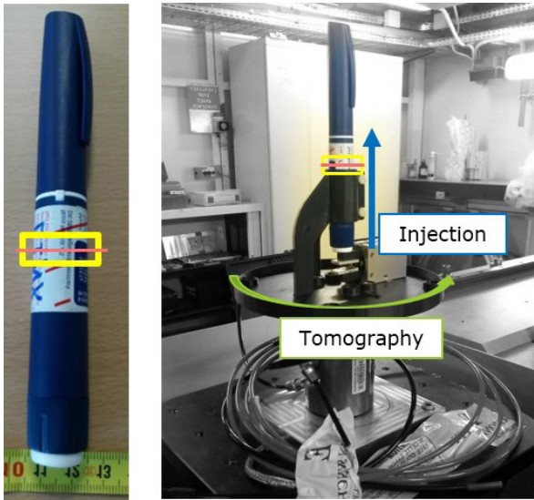
Insulin injection pens consist of many moving parts that have to work in unison to deliver the exact dosis of insulin to the patient subdermally. To capture the very fast movements of the internal parts, time-resolved imaging is performed. For such fast CT acquisitions, synchrotron radiation facilities offer CT scans with sub-second time resolution, acquiring several full tomograms in one second. However, even for the acquisition of 3 tomograms per second, the real movements still had to be slowed down. In addition to the time resolution challenge, the typical field of view used at synchrotron radiation sources is small, limiting the area of the insulin pen that can be visualized.
Collaboration
Members of the 3D Imaging Center at DTU travelled together with one representative of Novo Nordisk to the European Synchrotron Radiation Facility in Grenoble, France to record high-resolution X-ray tomography scans at the microtomography beamline ID 19 on insulin injection pens during dose application. The visualization and postprocessing of the CT data was carried out by DTU. This collaboration was part of the LINX project, in which researchers at leading Danish universities collaborate with scientists in industry to solve industry relevant problems using advanced neutron and X-ray techniques.
Results
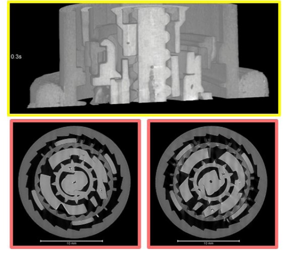
The insulin pen was set up at the beamline ID 19 of ESRF to be remotely activated while 53 X-ray tomography scans were taken continuously during its operation. Different positions of the pen were studied while modifying the injection speed. At a time resolution of 3 full tomograms per second, the tomography acquisition was performed with a spatial resolution of 24 µm, which allowed to see the movements of the internal parts with decent spatial and temporal resolution.
Perspectives
Further data treatment is required to improve the image quality for better visualizations. This is performed in collaboration with the QIM Center at DTU, a part of the 3D Imaging Center at DTU.