Optimizing X-ray imaging for fast data acquisition

Challenge
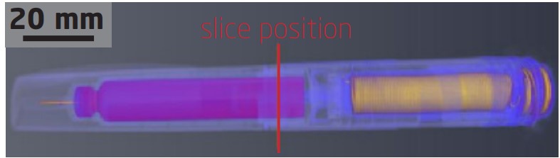
An insulin injection pen consists of many small moving parts which can be visualised by X-ray micro CT. In order to operate at a high speed, both the acquisition parameters and the reconstrution need to be optimised. This can be done by varying e.g. how many individual X-ray images are taken while the object is rotated (“number of projections”) and by how long the detector is illuminated by the X-ray beam (“integration time”). However, when using few projections and the common reconstruction method based on a “filtered back projection”, the quality of the reconstructed slices is usually negatively affected by stripes in the images. Using e.g. the “Simultaneous Iterative Reconstruction Technique” (SIRT) instead - a reconstruction algorithm method for correcting artifacts, such as stripes originating from from few projections - the resolution and contrast of the reconstructed data from CT scans with limited amounts of projections can be optimised.
Collaboration
This collaboration was part of the LINX project, in which researchers at leading Danish universities collaborate with scientists in industry to solve industry relevant problems using advanced neutron and X-ray techniques.
Results
Measurements were performed using a Nikon XT H 225 X-ray tomography setup. It was shown that a longer integration time and a higher number of projections resulted in a better quality of the reconstructed CT slices. While a filtered back projection-based reconstruction resulted in a good image quality from 1571 to 150 projections, the use of SIRT reconstruction could lower the number of projections to 78, which increases the scanning speed even more. For this reconstruction approach, an open reconstruction framework (Astra toolbox) was used and optimized accordingly.
Perspectives
Though the number of projections was considerably reduced, more powerful X-ray facilities are required to record the fast movements within the insulin pen in real time. These were performed at a later stage of this project at the European Synchroton Radiation Facility (ESRF) in France.